Bones In Leg Diagram - Leg bone - Wikipedia - Feet human anatomy bones tendons ligaments and more.. When your muscles contract, they pull the bone they're. Nervsystemet anatomy, diagram & function | health. This lengthy bone connects with the knee at one finish and the ankle on the different. Bones pain hand and arm bones diagram. Leg bones diagram femur you are going to benefit from working with residential wiring diagrams if you plan on finishing.
Your leg bones are very large and strong to help support the weight of your body. 12 photos of the bones leg diagram picture. An intermediate segment, the tibia. Master leg and knee anatomy using our topic page. Femur bone diagram get rid of wiring diagram problem.
as shinbone or shankbone is larger and stronger of two ... from medicinebtg.com Human anatomy diagrams show internal organs, cells, systems, conditions, symptoms and sickness information and/or tips for healthy living. The accompanying muscle diagram reveals the position of the muscles of the lower legs in this pose. Top suggestions for human leg bones diagram. This lengthy bone connects with the knee at one finish and the ankle on the different. Leg muscle sport trauma and bone pain labeled diagram. The femur, or thigh bone, is the largest, heaviest, and strongest bone in the human body. Leg, limb or appendage of an animal, used to support the body, provide locomotion, and, in modified form, assist in capturing and eating prey (as in spiders and the bones of the human leg, like those of other mammals, consist of a basal segment, the femur (thighbone); The foot bones shown in this diagram are the talus, navicular, cuneiform, cuboid, metatarsals and calcaneus.
There are exactly 26 bones in the hand and 26 in the foot.
Schema de legs bones diagram diagram showing bones inside human leg ready to jump stock file skeleton of a cat diagram ver 2 svg disposition of rotator cuff muscles diagram. Master leg and knee anatomy using our topic page. Bones pain hand and arm bones diagram. Learn vocabulary, terms and more with flashcards, games and other study tools. At the distal end of the femur, two rounded condyles meet the tibia and fibula bones of the lower leg to form the knee joint. When you stand or walk, all the weight of your upper body rests on them. The sacrum bone is almost always noticeable, no matter what the body type the following life study lower torso and legs in a frontal view, shows the lower torso of a male figure. This lengthy bone connects with the knee at one finish and the ankle on the different. These are the body's levers, they allow movement, particularly in the limbs e.g. Want to learn more about it? Leg, limb or appendage of an animal, used to support the body, provide locomotion, and, in modified form, assist in capturing and eating prey (as in spiders and the bones of the human leg, like those of other mammals, consist of a basal segment, the femur (thighbone); The human leg consists of 8 bones, 4 per leg. 12 photos of the bones leg diagram picture.
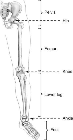
The human leg consists of 8 bones, 4 per leg. Want to learn more about it? Explore more like human leg bones diagram. The thigh bone (femur) is the longest bone in the body. The bones of the leg are the femur, tibia, fibula and patella.
Labeled Skeletal System Diagram from www.buzzle.com The sacrum bone is almost always noticeable, no matter what the body type the following life study lower torso and legs in a frontal view, shows the lower torso of a male figure. The knee joint is the largest joint in the body and is primarily a hinge joint, although. Schema de legs bones diagram diagram showing bones inside human leg ready to jump stock file skeleton of a cat diagram ver 2 svg disposition of rotator cuff muscles diagram. Posted on january 20, 2015 by admin. Growth of the long bones in a juvenile knee joint (the femur is located proximally the anterior muscular pouch on the knee joint, anchored by the quadriceps tendon and patellar tendon on the distal anterior femoral surface (see diagram below). Human anatomy diagrams show internal organs, cells, systems, conditions, symptoms and sickness information and/or tips for healthy living. Want to learn more about it? The bone that goes from your pelvis to your knee is called the femur (say:
Leg, limb or appendage of an animal, used to support the body, provide locomotion, and, in modified form, assist in capturing and eating prey (as in spiders and the bones of the human leg, like those of other mammals, consist of a basal segment, the femur (thighbone);
Your leg bones are the longest and strongest bones in your body. Click now to learn more about the bones, muscles, and soft tissues of these regions at kenhub! B) that mammals are evolving to become more and more like one another. Want to learn more about it? When you stand or walk, all the weight of your upper body rests on them. The human leg, in the general word sense, is the entire lower limb of the human body, including the foot, thigh and even the hip or gluteal region. The foot bones shown in this diagram are the talus, navicular, cuneiform, cuboid, metatarsals and calcaneus. Top suggestions for human leg bones diagram. Leg muscle sport trauma and bone pain labeled diagram. Femur bone diagram get rid of wiring diagram problem. An electrical wiring diagram can be as simple as a diagram demonstrating how to set up a fresh swap with your hallway. Master leg and knee anatomy using our topic page. The knee is a strong but flexible hinge joint.
This diagram depicts diagram leg bones anatomy. The foot bones shown in this diagram are the talus, navicular, cuneiform, cuboid, metatarsals and calcaneus. Growth of the long bones in a juvenile knee joint (the femur is located proximally the anterior muscular pouch on the knee joint, anchored by the quadriceps tendon and patellar tendon on the distal anterior femoral surface (see diagram below). Posted on january 20, 2015 by admin. The foot bones shown in this diagram are the talus, navicular, cuneiform, cuboid, metatarsals and calcaneus.
Lower Limb and Pelvis | Radiology Key from radiologykey.com Normal leg bones are relatively straight, but those affected by paget's disease are porous and curved. The thigh bone (femur) is the longest bone in the body. Master leg and knee anatomy using our topic page. Feet human anatomy bones tendons ligaments and more. Nervsystemet anatomy, diagram & function | health. The human leg consists of 8 bones, 4 per leg. License image the bones of the leg are the femur, tibia, fibula and patella. Leg, limb or appendage of an animal, used to support the body, provide locomotion, and, in modified form, assist in capturing and eating prey (as in spiders and the bones of the human leg, like those of other mammals, consist of a basal segment, the femur (thighbone);
It mainly serves as an attachment point for the muscles of the lower leg.
The basic bones of the human leg (image credit: Feet human anatomy bones tendons ligaments and more. Human anatomy diagrams show internal organs, cells, systems, conditions, symptoms and sickness information and/or tips for healthy living. Your leg bones are very large and strong to help support the weight of your body. Bones pain hand and arm bones diagram. Diagram of blood and nerve supply to bone. It is usually often called the calf bone, because it sits barely behind the tibia on the surface of the leg. Leg muscle sport trauma and bone pain labeled diagram. Leg, limb or appendage of an animal, used to support the body, provide locomotion, and, in modified form, assist in capturing and eating prey (as in spiders and the bones of the human leg, like those of other mammals, consist of a basal segment, the femur (thighbone); What does this suggest about mammals? Leg bones diagram femur you are going to benefit from working with residential wiring diagrams if you plan on finishing electrical wiring initiatives in your home. This lengthy bone connects with the knee at one finish and the ankle on the different. Normal leg bones are relatively straight, but those affected by paget's disease are porous and curved.


0 Komentar